Month: October 2019

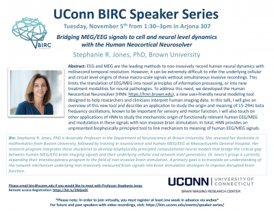
Talk: Stephanie Jones, Brown University

Brown University

Tuesday, November 5th from 1:30-3:00 pm in Arjona 307

Abstract: EEG and MEG are the leading methods to non-invasively record human neural dynamics with millisecond temporal resolution. However, it can be extremely difficult to infer the underlying cellular and circuit level origins of these macro-scale signals without simultaneous invasive recordings. This limits the translation of EEG/MEG into novel principles of information processing, or into new treatment modalities for neural pathologies. To address this need, we developed the Human Neocortical Neurosolver (HNN: https://hnn.brown.edu), a new user-friendly neural modeling tool designed to help researchers and clinicians interpret human imaging data. In this talk, I will give an overview of this new tool and describe an application to study the origin and meaning of 15-29Hz beta frequency oscillations, known to be important for sensory and motor function. I will also touch on other applications of HNN to study the mechanistic origin of functionally relevant human EEG/MEG and modulation in these signals with non-invasive brain stimulation. In total, HNN provides an unpresented biophysically principled tool to link mechanism to meaning of human EEG/MEG signals.

Bio: Stephanie R. Jones, PhD is Associate Professor in the Department of Neuroscience at Brown University. She received her doctorate in mathematics from Boston University, followed by training in neuroscience and human MEG/EEG at Massachusetts General Hospital. Her research program integrates these disciplines to develop biophysically principled computational neural models that bridge the critical gap between human MEG/EEG brain imaging signals and their underlying cellular and network level generators. Dr. Jones’s group is currently expanding their interdisciplinary program to the field of non-invasive brain stimulation. A primary goal is to translate an understanding of the network mechanism underlying non-invasively measured brain signals into brain stimulation strategies to improve disrupted brain function.

**To view this talk remotely via Webex, please register hereby October 29th**

Please email birc@uconn.edu if you are interested in meeting with a speaker. Click here to see the full BIRC Speaker Series schedule and access recordings of past talks.
Inspirational Faculty Presentation: Can Neuroscience Democratize Education?

UConn BIRC Director, Fumiko Hoeft, gave one of the “Inspirational Faculty Presentations” at UConn President’s Inauguration on October 4th, 2019. In her talk, she discussed how innovation in neuroscience science can democratize education and practice. Recording of all presentation, including Fumiko Hoeft’s, can now be viewed here.

MRI Scanner Operation Training for Qualified Candidates


The Brain Imaging Research Center now offers qualified candidates the opportunity to learn how to operate the Siemens Prisma 3T MRI Scanner to perform brain research studies. This training will consist of three components:
Didactic – All candidates will be required to attend a two-hour class about MRI safety tailored to issues that can be encountered during data acquisition. This training will include: Preventing radiofrequency (RF) burns; working with Specific Absorption Rates (SAR); proper participant preparation; quench emergency procedures. Note: a basic knowledge of MRI physics is necessary for this class.
Instrumentation – All candidates will be required to attend a two-hour class to learn basic scanner operation, including: User platform orientation (Syngo VE11C); coil selection and handling; participant positioning; BOLD screen operation; Eye Link operation; response box selection and operation; image transfer to NiDB or XNAT; basictroubleshooting.
Scanner Operation – All candidates will be required to successfully complete a minimum of twenty research scan sessions that include fMRI (BOLD) imaging, structural sequences, and DTI (diffusion tensor imaging) with direct supervision by an MRI Technologist. Scanning studies currently active at BIRC may complete this requirement. Additional scan sessions may be required at the discretion of the supervising technologist.
After successful completion of the above training, the candidate will be allowed to scan his or her own studies without direct supervision. Note: An MRI technologist must be in the facility for all scan sessions.
Candidates must maintain their status by completing a minimum of one study per month. Failure to do so will require a minimum of two directly supervised scan sessions per missed month.
Training is expected to begin the week of October 28 2019, allowing the candidate to be prepared to scan his or her own study during the spring semester.
Qualified candidate prerequisites:
      • Post doc with a commitment to remain for a minimum of one year (must be endorsed by PI)
      • Graduate student who has completed their Masters degree and must be endorsed by PI
      • Formal knowledge of basic MRI physics
      • Completion of Level 1 and Level 2 Safety Training
      • CPR certified (must provide documentation prior to scanning humans)

Online classes available at redcross.org/take-a-class/online-safety-classes

Interested candidates can apply for this training opportunity by providing the following information and documentation:
      • PI name, duration of contract, and written endorsement
      • Proof of formal basic MRI physics education
      • Any previous MRI experience
      • Study name, projected start date, and expected number of participants
Important dates:
      • Application submission: October 7-October 18 2019
      • Candidate acceptance notification: October 25 2019
      • Didactic and Instrumentation training: November 2019 (dates TBD)
      • Scanner Operation: November 2019 until completed
Please send the requested information to:
 

Elisa Medeiros, R.T.(R)(CT)(MR)
MRSO (MRSC™)
Manager, MRI Services
University of Connecticut
Brain Imaging Research Center
2 Alethia Drive Unit 1271
Storrs CT 06269-1271