There is a scheduled power shutdown on South Campus starting Saturday 10/13 at 8PM and ending Sunday 10/14 at 8PM. The BIRC facility will not be accessible during the shutdown. The NiDB, wiki, and scheduler systems will not be affected.
Uncategorized
Talk: Functional MRI Studies of Memory and Navigation
Chantal Stern, D.Phil
Boston University, Psychological and Brain Sciences
Wednesday, September 19 2018 3:30-4:30PM in BOUS A106
Biography:
Stern is an expert in human brain imaging and was a member of the research team that pioneered the development of functional magnetic resonance imaging, including early work focusing on the human hippocampus. Her lab’s primary goal is to study how the normal brain encodes, stores, and subsequently recognizes visual, spatial, and verbal information. In addition to studies of normal memory processes, including long-term and short-term memory processes, Stern and her team are studying basic science questions that include understanding spatial navigation, rule-learning, and interactions between memory and attention. Her translational work focuses on Alzheimer’s and Parkinson’s diseases. Thanks to a $1.6 million National Science Foundation instrumentation grant that Stern secured in 2016, her center showcases a Siemens 3 Tesla Magnetic Resonance Imaging scanner—a fundamental tool for studying the human brain. (Biography courtesy of www.bu.edu)
Vistors from UCHC are encourage to use the UCHC-Storrs shuttle service. Talks can also be joined remotely. Please contact us if you are interested in meeting with the speaker.
Talk: Neuroimaging Markers of Cognitive Reserve and Brain Aging
Lihong Wang PhD
UCONN Health, Dept of Psychiatry
Wednesday September 5, 2018 1:30-2:30pm in Arjona 307
Abstract
Our brain can reorganize its function and neural resources to counteract neural damages. The ability of reorganization of brain function depends on cognitive reserve capacity. To examine dynamic changes of cognitive reserve over time, we developed a new measure for evaluating neural compensatory capacity, a core factor of cognitive reserve, using independent component analysis and a cognitively very challenging task in older adults. Interestingly, we find higher neural compensatory capacity to be related to working memory function. In another study, we show a one-month physical exercise training to improve working memory as well as neural compensatory capacity through activating addition neural networks, i.e., the cerebellar and motor cortex. We believe the new measure on neural compensatory capacity can be applied to broad lines of research on neuroplasticity. Other imaging markers related to brain aging and cognitive decline will also be discussed.
Speaker
Dr. Wang obtained her Ph.D. degree in neurology from Japan and has six years of experience as a neurologist in China. She has performed neuroimaging-related research in depression at Duke University for over 12 years, primarily focused on geriatric depression and cognitive neuroscience. Her recent research centers on neural signatures of depression vulnerability and neural plasticity in patients with late-life depression and mild cognitive decline.
Vistors from UCHC are encourage to use the UCHC-Storrs shuttle service. Talks can also be joined remotely. Please contact us if you are interested in meeting with the speaker.
Introducing the new Scientific Director (August 2018)
The BIRC is delighted to welcome our new Scientific Director: Dr. Fumiko Hoeft, M.D./ Ph.D. She is a cognitive neuroscientist, with theoretical interests in the neurobiological mechanisms underlying individual differences in brain maturational processes and the acquisition of skills such as reading (and dyslexia). She will be leading the BIRC, and joining the Department of Psychological Sciences. She brings an impressive track record of externally-funded research and development, and a dynamic vision for the future of BIRC. Welcome, Dr. Hoeft!

Talk: Stephen Wilson 4/4 3:30
Stephen Wilson, PhD
Vanderbilt
Wednesday, April 4, 2018
3:30-5:00pm
BOUS A106
Imaging the language network: functional neuroanatomy, acquired aphasia, and recovery
What is the functional architecture of the language network? How is it impacted by damage to its various nodes and connections? And when it is damaged, how can it reorganize to support recovery of language function? To address these questions, we have carried out a series of multimodal neuroimaging studies in individuals with acquired language deficits of diverse etiologies–stroke, neurodegenerative disease, and resective surgery–as well as neurologically normal volunteers. Our findings, along with those of others, reveal a complex, variegated language network in which numerous distinct regions and tracts in the temporal, frontal and parietal lobes play distinct functional roles. Yet the network is strikingly resilient to most patterns of damage, indicating that in many cases, functional specialization is graded rather than absolute. Our findings suggest that recovery from aphasia depends primarily on reconfiguration of spared language regions, rather than macroscopic reorganization of the whole system.
Talk: Evelina Fedorenko 3/28 3:30pm
Evelina Fedorenko, PhD
Assistant Professor
Harvard Medical School and Massachusetts General Hospital
Wednesday March 28, 2018
3:30-5:00pm, BOUS A106
The cognitive and neural architecture of the human language system
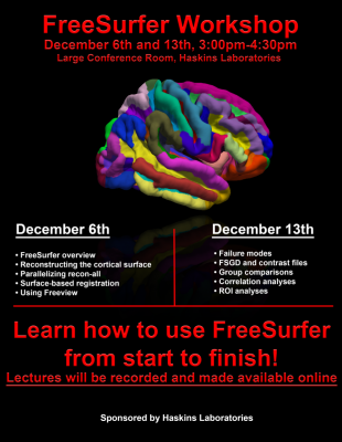
Interested in analyzing structural MRI data? Freesurfer workshop, 12/6-12/13

New England Research on Dyslexia Society Meeting October 21
The 3rd meeting of the New England Research on Dyslexia Society will be held in Storrs, CT on October 21, 2017. The meeting will take place on the University of Connecticut campus in Oak Hall.
KEYNOTE SPEAKER: John Gabrieli, Ph.D, Professor of Brain and Cognitive Sciences, MIT
The New England Research Group on Dyslexia is an interdisciplinary community of researchers, educators, clinicians, and policy experts, whose work aims at elucidating the biological, including psychological, and social underpinnings of Developmental Dyslexia and related disorders with the objective of improving prevention, early detection, diagnosis, treatment/intervention and social support (including legal, political, and public health) associated with this learning disability.
Registration is closed.
BOLD Brownbag Series
BIRC is pleased to present a series of informal BOLD Brownbag talks, held on Wednesdays from 9-10 AM in Bous 162. If you are not currently on the MRI distribution list, but would like to hear updates about these talks, please join the list serve. This venue is an informal one, geared toward discussion of work in progress (especially methodological/analytical) rather than formal polished presentations of finished products (though we’ll have some of those as well!).
Change in leadership at BIRC
After serving as Director since BIRC’s inception, Jay Rueckl will taking a well-earned sabbatical leave. Inge-Marie Eigsti will step in as Acting Director of BIRC. Dr. Eigsti’s imaging research centers on neural circuitry underlying the processing of language and special interests in autism spectrum disorder (see a recent article here).