Expanding Minds: BIRC Community Outreach

The UConn BIRC was recently featured in UConn Today for community outreach. The center hosted students from E.O. Smith High School for a tour of the facility and informational sessions led by faculty, staff, and graduate students.

For the original story in UConn Today and photographs, please click here.

Fumiko Hoeft Receives Eye-to-Eye Academic Excellence Award

Fumiko Hoeft, MD, PhD recently received an award from nonprofit organization Eye-to-Eye for her work with Stephanie Haft: Impact of mentoring on socio‐emotional and mental health outcomes of youth with learning disabilities and attention‐deficit hyperactivity disorder. The paper can be accessed here.

To learn more about Eye-to-Eye and their mission, visit their website.

InCHIP Virtual Meet ‘n’ Greet: UConn Brain Imaging Imaging Research Center

Dr. Fumiko Hoeft, Director of the UConn Brain Imaging Research Center (BIRC) shares information regarding the state-of-the-art equipment, methods, and training offered by BIRC and how the center supports both brain and whole-body imaging and research across the life span in addition to a range of clinical and nonclinical populations. She also covers BIRC equipment that can be used for research purposes includes MRI/fMRI scanner, TMS, tDCS/tACS, and EEG.

Also, watch to learn more about a $30,000 seed grant opportunity that InCHIP and BIRC are co-sponsoring!

Click here to watch the full InCHIP Virtual Meet ‘n’ Greet BIRC Seminar

Click here to view the slide deck in pdf format

Talk: Michele Diaz, Penn State

Michele Diaz, Ph.D.

Pennsylvania State University

Wednesday, May 1st from 1:30-2:30 in Arjona 307

Abstract: Although decline in cognitive functions is often observed with aging, language functions show a pattern of both impaired and spared performance. Semantic processes, such as vocabulary, are well maintained throughout adulthood. In contrast, older adults show impairments in phonological aspects of language production such as in increased slips of the tongue and increased pauses during speech. This asymmetric pattern suggests a fundamental difference in the cognitive and neural organization of these two language abilities. In this talk, I will discuss our work which has looked at semantic and phonological decisions, as well as more recent work that has examined inherent aspects of words, such as phonological and semantic neighborhood densities. Our work examines how cognition, behavior, and neural factors relate to each other and how they contribute to language function in healthy younger and older adults.

Bio: Professor Michele Diaz, PhD is currently Associate Professor of Psychology and Linguistics as well as Director of Human Imaging, at the Social, Life, & Engineering Sciences Imaging Center (SLEIC) at Pennsylvania State University. Previously, she has used electrophysiology to examine how semantic and phonological processes interact during spoken language comprehension. Using functional Magnetic Resonance Imaging (fMRI), she has investigated the neural substrates of orthographic, lexical, and semantic aspects of visual word processing, and the influence of conscious perception on the engagement of brain regions that support these processes. Finally, her involvement with the Biomedical Informatics Research Network (BIRN) project has allowed her to explore technical issues related to multi-site imaging, such as quality assurance measures and reliability. This experience facilitates her current role as the Director of Human Imaging at the Social, Life, and Engineering Sciences Imaging Center. 

Visitors from UCHC are encouraged to use the UCHC-Storrs shuttle service. Talks can also be joined remotely. Please contact us if you are interested in meeting with the speaker.


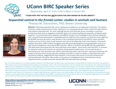
Talk: Dr. Theresa Desrochers, Brown University

Theresa M. Desrochers, PhD

Brown University

Wednesday, April 3rd from 1:30-2:30 in Arjona 307

Abstract: Performing sequential tasks such as making your breakfast are an integral part of daily life. The majority of previous studies have focused on motor sequences or non-sequential abstract control, rather than these kinds of more abstract sequential tasks. Our work using high-density multi-electrode chronic recordings in nonhuman primates has shown that an integrated cost-benefit signal in the striatum predicts the acquisition of habitual motor sequences. To move beyond motor sequences and address this gap in our knowledge of more abstract sequential tasks, we asked human participants to repeatedly perform simple four-item sequences of shape and color judgments during fMRI scanning. We found a novel dynamic in the rostrolateral prefrontal cortex (RLPFC), where activation ramped up across the four items in each sequence and reset at the beginning of each new sequence. Transcranial magnetic stimulation (TMS) to RLPFC during the same task selectively produced an increasing pattern of errors as each sequence progressed, mirroring the fMRI activation. Effects in the RLPFC during fMRI and two independent TMS experiments dissociated from two other prefrontal control regions. These results show that RLPFC is necessary for sequential control and resolution of uncertainty during sequence performance. Current work focuses on dissociating some of the processes that underlie sequential task control: task execution, sequential monitoring, and sequence memory. Recent results show that ramping in the RLPFC is robust to changes in sequential stimuli and monitoring conditions, suggesting that these dynamics in the frontal cortex may be a common mechanism for tracking sequential information. New studies are focusing on investigating frontal cortical dynamics during sequential control in parallel nonhuman primate fMRI and multi-electrode recordings.

Bio: Dr. Theresa Desrochers is currently Assistant Professor in the Departments of Neuroscience and Psychiatry & Human Behavior at Brown University. She earned her PhD in Neuroscience from the Massachusetts Institute of Technology in 2011. There she trained with Dr. Ann M. Graybiel and co-developed a new method of performing high-density, reconfigurable recordings on awake-behaving nonhuman primates. For her postdoctoral fellowship, Dr. Desrochers worked with Dr. David Badre at Brown University where she discovered a novel brain dynamic that was necessary for the sequential executive functions. Dr. Desrochers joined the faculty of the Department of Neuroscience at Brown University in the fall of 2016. The Desrochers lab uses human and nonhuman primate models to investigate the neural underpinnings of sequential control. Work in the lab focuses on explicitly addressing these questions using a cross-species approach, which is rare in both human and nonhuman primate research. Current experiments are focused on using nonhuman primate fMRI, a technique that only a few labs are able to use, to explicitly bridge between human fMRI and nonhuman primate neural recordings and directly examine functional homology between the species.

Visitors from UCHC are encouraged to use the UCHC-Storrs shuttle service. Talks can also be joined remotely. Please contact us if you are interested in meeting with the speaker.

Call for InCHIP-BIRC Seed Grants

UConn’s Institute for Collaboration on Health, Intervention, and Policy (InCHIP) and the UConn Brain Imaging Research Center (BIRC) are co-sponsoring a seed grant funding opportunity for faculty at UConn Storrs, UConn Health, and the regional campuses. This grant is designed to fund an innovative pilot project that will directly support an external grant application in health behavior or health policy with a substantial neuroimaging component. Health is broadly defined and includes physical and mental health and outcomes with critical implications for health. The funded pilot project must involve neuroimaging- related research that includes MRI, TMS, tDCS/tACS, and/or EEG usage at BIRC.

One seed grant of $30,000 is available through this seed grant competition. Funding in the amount of $15,000 (half of the seed grant) will be provided in the form of a limited number of allocated hours on MRI, EEG, and/or TMS equipment at BIRC. The remaining $15,000 may be used to fund other research costs associated with the pilot project.

This funding is intended to enable investigators to demonstrate feasibility, develop scientific and technical expertise, establish collaborations, and, secondarily, publish in peer-reviewed journals.

One of the goals of this funding mechanism is to encourage incorporation of BIRC’s neuroimaging services into the pilot project. Therefore, at least one of the Principle Investigators on the seed grant application must be new to BIRC and not have previously utilized BIRC’s neuroimaging services.

Investigators with experience in neuroimaging methods, as well as those with little or no experience may apply for this grant, but those with limited experience must include a collaborative plan for how they will acquire such experience.

An external grant application should be submitted through InCHIP within one year of completing the pilot project.

Key Dates

  • Friday, February 15, 2019 FOA posted
  • Monday, April 8, 2019 12-12:45pm optional webinar (email boundaryspanners@chip.uconn.edu to reserve)
  • Friday, April 26, 2019 Letters of Intent (required) due by 11:59 PM EST
  • Friday, May 3, 2019 Applicants notified of LOI approval decision
  • Friday, May 17, 2019 InCHIP Affiliate Application due by 11:59 PM EST
  • Friday, May 31, 2019 Full Proposals due by 11:59 PM EST
  • June 2019 Applicants notified of award decision
  • July 1, 2019 – June 30, 2021 Award period

To Apply

Click here for the full FOA and application form.

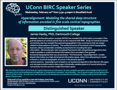
Talk: Dr. James V. Haxby, Dartmouth College

James V. Haxby, PhD

Dartmouth College

Distinguished Speaker

Wednesday, February 20 2019 3:30-5:00PM Bousfield A106

Abstract: Multivariate pattern analysis (MVPA) has revealed that information is encoded in finegrained patterns of cortical activity that can be measured with fMRI. Study of cortical functional connectivity also has revealed fine-grained topographies in the connectome that are closely related to these patterns of activity. The surface structure of functional cortical topographies, however, allows considerable variability across brains for encoding the same information. We introduced a new conceptual framework with computational algorithms that make it possible to model the shared information that is encoded in fine-grained functional topographies that vary across brains. This framework, “hyperalignment”, models shared information as a high-dimensional information space, rather than attempting to model a shared or canonical topographic structure in the physical space of cortical anatomy. Hyperalignment is based on computational algorithms that discover this space and calculate transformations that project individually-variable patterns of neural activity and connectivity into the common model information space.

Research Focus: My current research focuses on the development of computational methods for building models of representational spaces. We assume that distributed population responses encode information. Within a cortical field, a broad range of stimuli or cognitive states can be represented as different patterns of response. We use fMRI to measure these patterns of response and multivariate pattern (MVP) analysis to decode their meaning. We are currently developing methods that make it possible to decode an individual’s brain data using MVP classifiers that are based on other subjects’ data. We use a complex, natural stimulus to sample a broad range of brain representational states as a basis for building high-dimensional models of representational spaces within cortical fields. These models are based on response tuning functions that are common across subjects. Initially, we demonstrated the validity of such a model in ventral temporal cortex. We are working on building similar models in other visual areas and in auditory areas. We also plan to investigate representation of social cognition using this same conceptual framework.


Visitors from UCHC are encouraged to use the UCHC-Storrs shuttle service. Talks can also be joined remotely. Please contact us if you are interested in meeting with the speaker.


UConn BIRC Trailblazer Award Announced

Issue Date

December 19, 2018

Background

Since the opening of the University of Connecticut (UConn) Brain Imaging Research Center (BIRC) in June 2015, there has been an increase and diversification of user-base, neuroimaging-related extramural grants, and neuroimaging expertise of students and faculty. However, there is still room for greater utilization of BIRC, which presents opportunities for BIRC to offer the resources to perform high-profile and neuroimaging-intensive research that other fully occupied imaging centers cannot offer.

Objective

The BIRC Trailblazer Award was created to allow research teams to perform cutting-edge research and/or perform research that will benefit the BIRC community at-large. The objective of the 2019 BIRC Trailblazer Award is to fund: (1) high-risk high-reward projects with exceptional innovation that lead to raising the visibility of UConn, College of Liberal Arts and Sciences (CLAS) and BIRC; and/or (2) projects that will benefit the BIRC community at-large (e.g. methods development). The project is intended to lead to high-profile peer-review publications, release of a public database, and/or work that is cited and utilized by large-number of UConn researchers in their grants and manuscripts. The project should also lead to large-scale and high-profile extramural grant applications shortly after the end of the funding period.


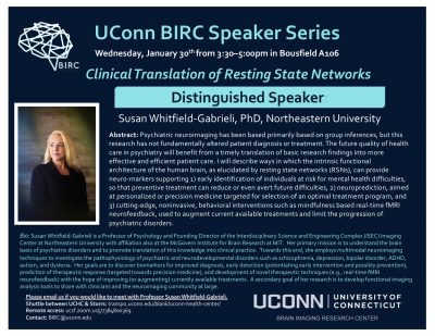
Talk: Clinical Translation of Resting State Networks

Susan Whitfield-Gabrieli, PhD

Northeastern University and MIT

Distinguished Speaker

Wednesday, January 30 2019 3:30-5:00PM Bousfield A106

Abstract: Psychiatric neuroimaging has been based primarily on group inferences, but this research has not fundamentally altered patient diagnosis or treatment. The future quality of healthcare in psychiatry will benefit from a timely translation of basic research findings into more effective and efficient patient care. I will describe ways in which the intrinsic functional architecture of the human brain, as elucidated by resting state networks (RSNs), can provide neuro-markers supporting 1) early identification of individuals at risk for mental health difficulties, so that perventive treatment can reduce or even avert future difficulties, 2) neuroprediction, aimed at personalized or precision medicine targeted for selection of an optimal treatment program, and 3) cutting-edge, noninvasive, behavioral interventions such as mindfulness based real-time fMRI neurofeedback, used to augment current available treatments and limit the progression of psychiatric disorders.

Bio: Susan Whitfield-Gabrieli is a Professor of Psychology and Founding Director of the Interdisciplinary Science and Engineering Complex (ISEC) Imaging Center at Northeastern University with affiliation also at the McGovern Institute for Brain Research at MIT. Her primary mission is to understand the brain basis of psychiatric disorders and to promote the translation of this knowledge into clinical practice. Towards this end, she employs multimodal neuroimaging techniques to investigate the pathophysiology of psychiatric and neurodevelopmental disorders such as schizophrenia, depression, bipolar disorder, ADHD, autism, and dyslexia. Her goals are to discover biomarkers for improved diagnosis, early detection (potentiating early intervention and possibly prevention), prediction of the therapeutic response (targeted towards precision medicine), and development of novel therapeutic techniques (e.g., real-time fMRI neurofeedback) with the hope of improving (or augmenting) currently available treatments. A secondary goal of her research is to develop functional imaging analysis tools to share with clinicians and the neuroimaging community at large.

Visitors from UCHC are encouraged to use the UCHC-Storrs shuttle service. Talks can also be joined remotely. Please contact us if you are interested in meeting with the speaker.