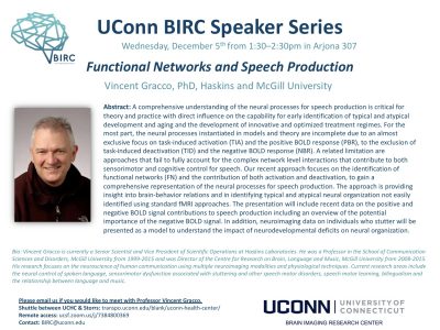
Clinical MRI scans now being offered at BIRC

UConn Health Patients Can Now Get MRIs at UConn in Storrs

MRI service manager Elisa Medeiros prepares a patient for functional MRI testing at the BIRC in the Phillips Communication Sciences Building. (Peter Morenus/UConn Photo)

UConn Health patients in eastern Connecticut will now be able to get MRI scans done in Storrs just as if they were at UConn Health in Farmington, thanks to a collaboration between doctors and researchers at the two campuses.

UConn’s Brain Imaging Research Center (BIRC) houses a powerful 3 Tesla Magnetic Resonance Imaging (MRI) scanner that was installed in 2015 and originally dedicated purely to research. The BIRC’s machine can take detailed pictures of fine structures in the brain. It can also do functional MRI, which shows subtle changes in blood flow in the brain as a person thinks. Medical details – tiny flecks of blood that might signal a concussion, or small injuries to the spine or extremities – also show up beautifully on the scanner. But the state had not previously licensed the BIRC’s machine to perform medical work. Doctors at UConn Health and researchers at the BIRC thought that should change.

“Soon after I started as chair, it became clear we had a long history of our UConn Husky athletes having scans done on the outside. But then their docs would bring the scans to us for a second read because they trusted us,” says Dr. Leo Wolansky, head of radiology at UConn Health. “It’s our moral obligation to take care of our own people,” but it was a lot of unpaid work too, he observes. Other patients in eastern Connecticut, including students, staff, and faculty, who wanted to use UConn Health doctors but worked and lived near Storrs also found it inconvenient to drive to Farmington for a scan. That meant a lot of UConn money leaving the institution, Wolansky adds, an undesirable phenomenon known as “leakage.”

So he began working with former BIRC scientific directors Inge-Marie Eigsti and Jay Rueckl, and more recently BIRC director Fumiko Hoeft, along with regulatory and business development staff at UConn Health, to get permission from the state to use the center’s machine for medical imaging. Then, with the help of UConn Health MRI technologist Brian Hausner, MRI service manager Elisa Medeiros installed protocols to run clinical scans. The picture archiving communication system team at UConn Health oversaw the setup of the hardware needed to transmit medical data securely from the BIRC, which is located in the Phillips Communication Sciences Building in Storrs, to UConn Health in Farmington.

It took months of work, but on Nov. 7, the BIRC scanned Clinical MRI Patient #1. The term “Clinical MRI Patient #1” had a double meaning. It referred to the first clinical scan chronologically, but it was also a HIPAA-compliant way to refer to the top executive at UConn, President Herbst, who agreed to be the “test case.” She has given permission to publish this information, and wants everyone to know she’s fine.

“The President’s support of the project was critical,” says Wolansky.

UConn Health doctors can now schedule MRIs for their patients at the BIRC in Storrs for Monday and Wednesday afternoons as easily as if they were going to the imaging center in Farmington. Urgent scans can be squeezed in at other times on a case-by-case basis. The BIRC capacity will free up some space at UConn Health, bringing new patients into the system, and is not expected to impact research done at the center at all.

“The biggest benefit is the integration between campuses. It’s a huge success for us to do this,” says Hoeft, the director of BIRC, noting that revenue from the scans will enhance the financial stability of the center.

Wolansky, who is based in Farmington, agrees. UConn’s Farmington and Storrs campuses function largely independently from one another, so this collaboration is based on goodwill, he says.

“They are UConn and we are UConn,” he adds. “Even though [patients] may be 40 minutes away by car, when we read the scans [at the imaging center in Farmington], it’s no different than if the patients were down the hall!”

This story was originally published in UConn Today


Talk: Functional Networks and Speech Production

Vincent Gracco, PhD

Wednesday December 5, 2018 1:30-2:30 PM Arjona 307

Haskins and McGill University

Abstract: A comprehensive understanding of the neural processes for speech production is critical for theory and practice with direct influence on the capability for early identification of typical and atypical development and aging and the development of innovative and optimized treatment regimes. For the most part, the neural processes instantiated in models and theory are incomplete due to an almost exclusive focus on task-induced activation (TIA) and the positive BOLD response (PBR), to the exclusion of task-induced deactivation (TID) and the negative BOLD response (NBR). A related limitation are approaches that fail to fully account for the complex network level interactions that contribute to both sensorimotor and cognitive control for speech. Our recent approach focuses on the identification of functional networks (FN) and the contribution of both activation and deactivation, to gain a comprehensive representation of the neural processes for speech production. The approach is providing insight into brain-behavior relations and in identifying typical and atypical neural organization not easily identified using standard fMRI approaches. The presentation will include recent data on the positive and negative BOLD signal contributions to speech production including an overview of the potential importance of the negative BOLD signal. In addition, neuroimaging data on individuals who stutter will be presented as a model to understand the impact of neurodevelopmental deficits on neural organization.

Bio: Vincent Gracco is currently a Senior Scientist and Vice President of Scientific Operations at Haskins Laboratories. He was a Professor in the School of Communication Sciences and Disorders, McGill University from 1999-2015 and was Director of the Centre for Research on Brain, Language and Music, McGill University from 2008-2015. His research focuses on the neuroscience of human communication using multiple neuroimaging modalities and physiological techniques. Current research areas include the neural control of spoken language, sensorimotor dysfunction associated with stuttering and other speech motor disorders, speech motor learning, bilingualism and the relationship between language and music.

Visitors from UCHC are encouraged to use the UCHC-Storrs shuttle service. Talks can also be joined remotely. Please contact us if you are interested in meeting with the speaker.

Tour the Brain: BIRC at the International Dyslexia Association 2018 Annual Conference


The BIRC is thrilled to announce its first community outreach event. UConn BIRC is partnering with the International Dyslexia Association (IDA) and Neuroscape to host a virtual reality tour of the brain. Join us in making neuroscience fun and accessible to the public!

The VR tours will take place at the IDA Massachusetts and Connecticut booth located in the exhibit hall at Foxwoods Resort and Casino  October 24th through 26th.

For additional information, including registration, please visit IDA’s website.


Talk: Linking Late-Life Depression, Cognitive Impairment, and Dementia

David C. Steffens, M.D., M.H.S.

UCONN Health, Department of Psychiatry

Wednesday, November 7 2018 1:30-2:30PM Arjona 307

Abstract: Depression in late life has been associated with cognitive decline and new onset of dementia. Recent studies have linked the presence of neuroticism with cognitive decline among older depressed adults. Structural Imaging studies in geriatric depression have found that hippocampal volume and burden of white matter hyperintensities are associated with cognitive outcomes. Functional magnetic resonance imaging (fMRI) has recently been used to study late-life depression. Preliminary findings of fMRI in older adults have examined the relationships of neuroticism and depression, identifying the medial prefrontal cortex as a key area in emotion regulation and susceptibility to depression.

Speaker Bio: Dr. Steffens has been funded by the NIH for over twenty-five years. His research focuses on links between late-life depression and subsequent cognitive decline and development of dementia. Assessments include structural and functional brain imaging, neuropsychological testing, blood work for peripheral markers and genetics, personality assessment, functional status, and measures of stress and social support.

Visitors from UCHC are encouraged to use the UCHC-Storrs shuttle service. Talks can also be joined remotely. Please contact us if you are interested in meeting with the speaker.

Tips You Won’t Get at Grant Writing Courses, 10/3 1:30-2:30pm

The second BIRC Speaker Series talk of the term will be this Wednesday 10/3 1:30-2:30 in Arjona 307. Please join us for this talk and the complete series!

Tips You Won’t Get at Grant Writing Courses: Presentation and discussions on NIH, NSF, foundation grants and philanthropy
with speakers Emily Myers PhD, James Magnuson PhD, Fumiko Hoeft MD PhD, and Rachel Marshall and Louis Bach from UConn Foundation.

Vistors from UCHC are encourage to use the UCHC-Storrs shuttle service. Talks can also be joined remotely.

Talk: Functional MRI Studies of Memory and Navigation

Chantal Stern, D.Phil

Boston University, Psychological and Brain Sciences

Wednesday, September 19 2018 3:30-4:30PM in BOUS A106

Biography:

Stern is an expert in human brain imaging and was a member of the research team that pioneered the development of functional magnetic resonance imaging, including early work focusing on the human hippocampus. Her lab’s primary goal is to study how the normal brain encodes, stores, and subsequently recognizes visual, spatial, and verbal information. In addition to studies of normal memory processes, including long-term and short-term memory processes, Stern and her team are studying basic science questions that include understanding spatial navigation, rule-learning, and interactions between memory and attention. Her translational work focuses on Alzheimer’s and Parkinson’s diseases. Thanks to a $1.6 million National Science Foundation instrumentation grant that Stern secured in 2016, her center showcases a Siemens 3 Tesla Magnetic Resonance Imaging scanner—a fundamental tool for studying the human brain. (Biography courtesy of www.bu.edu)

Vistors from UCHC are encourage to use the UCHC-Storrs shuttle service. Talks can also be joined remotely. Please contact us if you are interested in meeting with the speaker.

Talk: Neuroimaging Markers of Cognitive Reserve and Brain Aging

Lihong Wang PhD
UCONN Health, Dept of Psychiatry
Wednesday September 5, 2018 1:30-2:30pm in Arjona 307

Abstract

Our brain can reorganize its function and neural resources to counteract neural damages. The ability of reorganization of brain function depends on cognitive reserve capacity. To examine dynamic changes of cognitive reserve over time, we developed a new measure for evaluating neural compensatory capacity, a core factor of cognitive reserve, using independent component analysis and a cognitively very challenging task in older adults. Interestingly, we find higher neural compensatory capacity to be related to working memory function. In another study, we show a one-month physical exercise training to improve working memory as well as neural compensatory capacity through activating addition neural networks, i.e., the cerebellar and motor cortex. We believe the new measure on neural compensatory capacity can be applied to broad lines of research on neuroplasticity. Other imaging markers related to brain aging and cognitive decline will also be discussed.

Speaker

Dr. Wang obtained her Ph.D. degree in neurology from Japan and has six years of experience as a neurologist in China. She has performed neuroimaging-related research in depression at Duke University for over 12 years, primarily focused on geriatric depression and cognitive neuroscience. Her recent research centers on neural signatures of depression vulnerability and neural plasticity in patients with late-life depression and mild cognitive decline.

Vistors from UCHC are encourage to use the UCHC-Storrs shuttle service. Talks can also be joined remotely. Please contact us if you are interested in meeting with the speaker.

Download and post a flyer in your area.

Introducing the new Scientific Director (August 2018)

The BIRC is delighted to welcome our new Scientific Director: Dr. Fumiko Hoeft, M.D./ Ph.D. She is a cognitive neuroscientist, with theoretical interests in the neurobiological mechanisms underlying individual differences in brain maturational processes and the acquisition of skills such as reading (and dyslexia). She will be leading the BIRC, and joining the Department of Psychological Sciences. She brings an impressive track record of externally-funded research and development, and a dynamic vision for the future of BIRC.  Welcome, Dr. Hoeft!