Author: webdev

Talk: Stephanie Jones, Brown University

Brown University

Tuesday, November 5th from 1:30-3:00 pm in Arjona 307

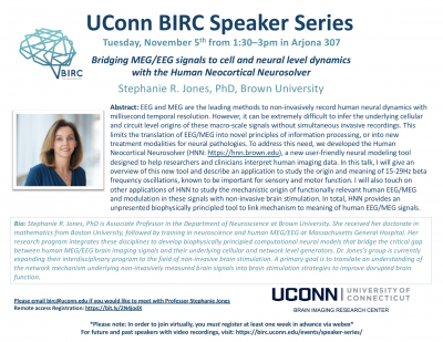
Abstract: EEG and MEG are the leading methods to non-invasively record human neural dynamics with millisecond temporal resolution. However, it can be extremely difficult to infer the underlying cellular and circuit level origins of these macro-scale signals without simultaneous invasive recordings. This limits the translation of EEG/MEG into novel principles of information processing, or into new treatment modalities for neural pathologies. To address this need, we developed the Human Neocortical Neurosolver (HNN: https://hnn.brown.edu), a new user-friendly neural modeling tool designed to help researchers and clinicians interpret human imaging data. In this talk, I will give an overview of this new tool and describe an application to study the origin and meaning of 15-29Hz beta frequency oscillations, known to be important for sensory and motor function. I will also touch on other applications of HNN to study the mechanistic origin of functionally relevant human EEG/MEG and modulation in these signals with non-invasive brain stimulation. In total, HNN provides an unpresented biophysically principled tool to link mechanism to meaning of human EEG/MEG signals.

Bio: Stephanie R. Jones, PhD is Associate Professor in the Department of Neuroscience at Brown University. She received her doctorate in mathematics from Boston University, followed by training in neuroscience and human MEG/EEG at Massachusetts General Hospital. Her research program integrates these disciplines to develop biophysically principled computational neural models that bridge the critical gap between human MEG/EEG brain imaging signals and their underlying cellular and network level generators. Dr. Jones’s group is currently expanding their interdisciplinary program to the field of non-invasive brain stimulation. A primary goal is to translate an understanding of the network mechanism underlying non-invasively measured brain signals into brain stimulation strategies to improve disrupted brain function.

**To view this talk remotely via Webex, please register hereby October 29th**

Please email birc@uconn.edu if you are interested in meeting with a speaker. Click here to see the full BIRC Speaker Series schedule and access recordings of past talks.

Inspirational Faculty Presentation: Can Neuroscience Democratize Education?

UConn BIRC Director, Fumiko Hoeft, gave one of the “Inspirational Faculty Presentations” at UConn President’s Inauguration on October 4th, 2019. In her talk, she discussed how innovation in neuroscience science can democratize education and practice. Recording of all presentation, including Fumiko Hoeft’s, can now be viewed here.

MRI Scanner Operation Training for Qualified Candidates


The Brain Imaging Research Center now offers qualified candidates the opportunity to learn how to operate the Siemens Prisma 3T MRI Scanner to perform brain research studies. This training will consist of three components:
Didactic – All candidates will be required to attend a two-hour class about MRI safety tailored to issues that can be encountered during data acquisition. This training will include: Preventing radiofrequency (RF) burns; working with Specific Absorption Rates (SAR); proper participant preparation; quench emergency procedures. Note: a basic knowledge of MRI physics is necessary for this class.
Instrumentation – All candidates will be required to attend a two-hour class to learn basic scanner operation, including: User platform orientation (Syngo VE11C); coil selection and handling; participant positioning; BOLD screen operation; Eye Link operation; response box selection and operation; image transfer to NiDB or XNAT; basictroubleshooting.
Scanner Operation – All candidates will be required to successfully complete a minimum of twenty research scan sessions that include fMRI (BOLD) imaging, structural sequences, and DTI (diffusion tensor imaging) with direct supervision by an MRI Technologist. Scanning studies currently active at BIRC may complete this requirement. Additional scan sessions may be required at the discretion of the supervising technologist.
After successful completion of the above training, the candidate will be allowed to scan his or her own studies without direct supervision. Note: An MRI technologist must be in the facility for all scan sessions.
Candidates must maintain their status by completing a minimum of one study per month. Failure to do so will require a minimum of two directly supervised scan sessions per missed month.
Training is expected to begin the week of October 28 2019, allowing the candidate to be prepared to scan his or her own study during the spring semester.
Qualified candidate prerequisites:
      • Post doc with a commitment to remain for a minimum of one year (must be endorsed by PI)
      • Graduate student who has completed their Masters degree and must be endorsed by PI
      • Formal knowledge of basic MRI physics
      • Completion of Level 1 and Level 2 Safety Training
      • CPR certified (must provide documentation prior to scanning humans)

Online classes available at redcross.org/take-a-class/online-safety-classes

Interested candidates can apply for this training opportunity by providing the following information and documentation:
      • PI name, duration of contract, and written endorsement
      • Proof of formal basic MRI physics education
      • Any previous MRI experience
      • Study name, projected start date, and expected number of participants
Important dates:
      • Application submission: October 7-October 18 2019
      • Candidate acceptance notification: October 25 2019
      • Didactic and Instrumentation training: November 2019 (dates TBD)
      • Scanner Operation: November 2019 until completed
Please send the requested information to:
 

Elisa Medeiros, R.T.(R)(CT)(MR)
MRSO (MRSC™)
Manager, MRI Services
University of Connecticut
Brain Imaging Research Center
2 Alethia Drive Unit 1271
Storrs CT 06269-1271

Talk: Uri Hasson, Princeton University

Uri Hasson, PhD

Princeton University

Tuesday, October 15th from 1:30-3:00 pm in Arjona 307

Abstract: Cognition materializes in an interpersonal space. At present, little is known about the neural substrates that underlie our ability to communicate with other brains in naturalistic settings. In the talk I will introduce novel methodological and analytical tools for characterizing the neural responses during production and comprehension of complex real-life speech. By directly comparing the neural activity timecourses during production and comprehension of the same narrative, we were able to identify areas in which the neural activity is correlated (coupled) across the speaker’s and listener’s brains during communication. Furthermore, the listener brain activity mirrors that of the speaker with a constant delay of three seconds. This mirroring was eliminated when the communication signals were misaligned. Finally, the stronger the speaker- listener coupling the greater listener comprehension. We argue that the observed coupling of production and comprehension-based processes serves as a mechanism by which brains convey information.

Bio: Uri Hasson grew up in Jerusalem. As an undergrad he studied philosophy and cognitive sciences at the Hebrew University. He completed his Ph.D. in Neurobiology at the Weizmann Institute in Israel and was a postdoctoral fellow at NYU before moving to Princeton. He is currently a Professor in the Psychology Department and the Neuroscience Institute at Princeton University. His research program aims to understand the neural basis of face-to-face, brain-to-brain, social interaction, with a focus on verbal communication and storytelling in real-life contexts.

**To view this talk remotely via Webex, please register here by October 8th**

Please email birc@uconn.edu if you are interested in meeting with a speaker. Click here to see the full BIRC Speaker Series schedule and access recordings of past talks.

Talk: Tor Wager, Dartmouth College

Tor Wager, PhD

Dartmouth College

Tuesday, September 10th from 1:30-3pm in Arjona 3o7

Abstract: Pain and emotion are central to human life. Their experience defines our wellbeing, and the brain processes that underlie them drive behavior and learning. Developing models of the brain systems that generate pain and emotion could transform how we understand their neurophysiological origins, and how we understand interventions ranging from drugs to psychotherapy. However, developing such models will require computational advances, particularly in our ability to model how emergent properties like pain arise from complex interactions among brain systems. In this talk, I describe an approach and a series of studies aimed at constructing models of pain and other forms of affect with high neuroscientific interpretability, predictive validity, and reproducibility. Combining fMRI with machine learning techniques, we have developed brain models capable of predicting the intensity of pain, negative affect, empathy, autonomic activity, and other subjective experiences in individual participants. In addition to utility as biomarkers and targets for both psychological and drug interventions, these models can provide insight into how the brain represents multiple varieties of affective experience. 

Bio: Tor Wager is the Diana L. Taylor Distinguished Professor in Neuroscience at Dartmouth College. He received his Ph.D. from the University of Michigan in Cognitive Psychology in 2003, and served as an Assistant (2004-2008) and Associate Professor (2009) at Columbia University, and as Associate (2010-2014) and Full Professor (2014-2019) at the University of Colorado, Boulder. Since 2004, he has directed the Cognitive and Affective Neuroscience laboratory, a research lab devoted to work on the neurophysiology of affective processes—pain, emotion, stress, and empathy—and how they are shaped by cognitive and social influences. Dr. Wager and his lab are also dedicated to developing analysis methods for functional neuroimaging and sharing ideas, tools, and scientific data with the scientific community and public. See http://wagerlab.colorado.eduand http://canlab.github.iofor papers, data, tools, and code.

**To view this talk remotely via Webex, please register here by September 3rd**

Please email birc@uconn.edu if you are interested in meeting with a speaker. Click here to see the full BIRC Speaker Series schedule and access recordings of past talks.

Expanding Minds: BIRC Community Outreach

The UConn BIRC was recently featured in UConn Today for community outreach. The center hosted students from E.O. Smith High School for a tour of the facility and informational sessions led by faculty, staff, and graduate students.

For the original story in UConn Today and photographs, please click here.

Fumiko Hoeft Receives Eye-to-Eye Academic Excellence Award

Fumiko Hoeft, MD, PhD recently received an award from nonprofit organization Eye-to-Eye for her work with Stephanie Haft: Impact of mentoring on socio‐emotional and mental health outcomes of youth with learning disabilities and attention‐deficit hyperactivity disorder. The paper can be accessed here.

To learn more about Eye-to-Eye and their mission, visit their website.

InCHIP Virtual Meet ‘n’ Greet: UConn Brain Imaging Imaging Research Center

Dr. Fumiko Hoeft, Director of the UConn Brain Imaging Research Center (BIRC) shares information regarding the state-of-the-art equipment, methods, and training offered by BIRC and how the center supports both brain and whole-body imaging and research across the life span in addition to a range of clinical and nonclinical populations. She also covers BIRC equipment that can be used for research purposes includes MRI/fMRI scanner, TMS, tDCS/tACS, and EEG.

Also, watch to learn more about a $30,000 seed grant opportunity that InCHIP and BIRC are co-sponsoring!

Click here to watch the full InCHIP Virtual Meet ‘n’ Greet BIRC Seminar

Click here to view the slide deck in pdf format

Talk: Michele Diaz, Penn State

Michele Diaz, Ph.D.

Pennsylvania State University

Wednesday, May 1st from 1:30-2:30 in Arjona 307

Abstract: Although decline in cognitive functions is often observed with aging, language functions show a pattern of both impaired and spared performance. Semantic processes, such as vocabulary, are well maintained throughout adulthood. In contrast, older adults show impairments in phonological aspects of language production such as in increased slips of the tongue and increased pauses during speech. This asymmetric pattern suggests a fundamental difference in the cognitive and neural organization of these two language abilities. In this talk, I will discuss our work which has looked at semantic and phonological decisions, as well as more recent work that has examined inherent aspects of words, such as phonological and semantic neighborhood densities. Our work examines how cognition, behavior, and neural factors relate to each other and how they contribute to language function in healthy younger and older adults.

Bio: Professor Michele Diaz, PhD is currently Associate Professor of Psychology and Linguistics as well as Director of Human Imaging, at the Social, Life, & Engineering Sciences Imaging Center (SLEIC) at Pennsylvania State University. Previously, she has used electrophysiology to examine how semantic and phonological processes interact during spoken language comprehension. Using functional Magnetic Resonance Imaging (fMRI), she has investigated the neural substrates of orthographic, lexical, and semantic aspects of visual word processing, and the influence of conscious perception on the engagement of brain regions that support these processes. Finally, her involvement with the Biomedical Informatics Research Network (BIRN) project has allowed her to explore technical issues related to multi-site imaging, such as quality assurance measures and reliability. This experience facilitates her current role as the Director of Human Imaging at the Social, Life, and Engineering Sciences Imaging Center. 

Visitors from UCHC are encouraged to use the UCHC-Storrs shuttle service. Talks can also be joined remotely. Please contact us if you are interested in meeting with the speaker.


Talk: Dr. Theresa Desrochers, Brown University

Theresa M. Desrochers, PhD

Brown University

Wednesday, April 3rd from 1:30-2:30 in Arjona 307

Abstract: Performing sequential tasks such as making your breakfast are an integral part of daily life. The majority of previous studies have focused on motor sequences or non-sequential abstract control, rather than these kinds of more abstract sequential tasks. Our work using high-density multi-electrode chronic recordings in nonhuman primates has shown that an integrated cost-benefit signal in the striatum predicts the acquisition of habitual motor sequences. To move beyond motor sequences and address this gap in our knowledge of more abstract sequential tasks, we asked human participants to repeatedly perform simple four-item sequences of shape and color judgments during fMRI scanning. We found a novel dynamic in the rostrolateral prefrontal cortex (RLPFC), where activation ramped up across the four items in each sequence and reset at the beginning of each new sequence. Transcranial magnetic stimulation (TMS) to RLPFC during the same task selectively produced an increasing pattern of errors as each sequence progressed, mirroring the fMRI activation. Effects in the RLPFC during fMRI and two independent TMS experiments dissociated from two other prefrontal control regions. These results show that RLPFC is necessary for sequential control and resolution of uncertainty during sequence performance. Current work focuses on dissociating some of the processes that underlie sequential task control: task execution, sequential monitoring, and sequence memory. Recent results show that ramping in the RLPFC is robust to changes in sequential stimuli and monitoring conditions, suggesting that these dynamics in the frontal cortex may be a common mechanism for tracking sequential information. New studies are focusing on investigating frontal cortical dynamics during sequential control in parallel nonhuman primate fMRI and multi-electrode recordings.

Bio: Dr. Theresa Desrochers is currently Assistant Professor in the Departments of Neuroscience and Psychiatry & Human Behavior at Brown University. She earned her PhD in Neuroscience from the Massachusetts Institute of Technology in 2011. There she trained with Dr. Ann M. Graybiel and co-developed a new method of performing high-density, reconfigurable recordings on awake-behaving nonhuman primates. For her postdoctoral fellowship, Dr. Desrochers worked with Dr. David Badre at Brown University where she discovered a novel brain dynamic that was necessary for the sequential executive functions. Dr. Desrochers joined the faculty of the Department of Neuroscience at Brown University in the fall of 2016. The Desrochers lab uses human and nonhuman primate models to investigate the neural underpinnings of sequential control. Work in the lab focuses on explicitly addressing these questions using a cross-species approach, which is rare in both human and nonhuman primate research. Current experiments are focused on using nonhuman primate fMRI, a technique that only a few labs are able to use, to explicitly bridge between human fMRI and nonhuman primate neural recordings and directly examine functional homology between the species.

Visitors from UCHC are encouraged to use the UCHC-Storrs shuttle service. Talks can also be joined remotely. Please contact us if you are interested in meeting with the speaker.