UConn BIRC Director, Fumiko Hoeft, gave one of the “Inspirational Faculty Presentations” at UConn President’s Inauguration on October 4th, 2019. In her talk, she discussed how innovation in neuroscience science can democratize education and practice. Recording of all presentation, including Fumiko Hoeft’s, can now be viewed here.
BIRC news
MRI Scanner Operation Training for Qualified Candidates
The Brain Imaging Research Center now offers qualified candidates the opportunity to learn how to operate the Siemens Prisma 3T MRI Scanner to perform brain research studies. This training will consist of three components:
Didactic – All candidates will be required to attend a two-hour class about MRI safety tailored to issues that can be encountered during data acquisition. This training will include: Preventing radiofrequency (RF) burns; working with Specific Absorption Rates (SAR); proper participant preparation; quench emergency procedures. Note: a basic knowledge of MRI physics is necessary for this class.
Instrumentation – All candidates will be required to attend a two-hour class to learn basic scanner operation, including: User platform orientation (Syngo VE11C); coil selection and handling; participant positioning; BOLD screen operation; Eye Link operation; response box selection and operation; image transfer to NiDB or XNAT; basictroubleshooting.
Scanner Operation – All candidates will be required to successfully complete a minimum of twenty research scan sessions that include fMRI (BOLD) imaging, structural sequences, and DTI (diffusion tensor imaging) with direct supervision by an MRI Technologist. Scanning studies currently active at BIRC may complete this requirement. Additional scan sessions may be required at the discretion of the supervising technologist.
After successful completion of the above training, the candidate will be allowed to scan his or her own studies without direct supervision. Note: An MRI technologist must be in the facility for all scan sessions.
Candidates must maintain their status by completing a minimum of one study per month. Failure to do so will require a minimum of two directly supervised scan sessions per missed month.
Training is expected to begin the week of October 28 2019, allowing the candidate to be prepared to scan his or her own study during the spring semester.
Qualified candidate prerequisites:
-
-
- Post doc with a commitment to remain for a minimum of one year (must be endorsed by PI)
- Graduate student who has completed their Masters degree and must be endorsed by PI
- Formal knowledge of basic MRI physics
- Completion of Level 1 and Level 2 Safety Training
- CPR certified (must provide documentation prior to scanning humans)
-
Online classes available at redcross.org/take-a-class/online-safety-classes
Interested candidates can apply for this training opportunity by providing the following information and documentation:
-
-
- PI name, duration of contract, and written endorsement
- Proof of formal basic MRI physics education
- Any previous MRI experience
- Study name, projected start date, and expected number of participants
-
Important dates:
-
-
- Application submission: October 7-October 18 2019
- Candidate acceptance notification: October 25 2019
- Didactic and Instrumentation training: November 2019 (dates TBD)
- Scanner Operation: November 2019 until completed
-
Please send the requested information to:
Elisa Medeiros, R.T.(R)(CT)(MR)
MRSO (MRSC™)
Manager, MRI Services
University of Connecticut
Brain Imaging Research Center
2 Alethia Drive Unit 1271
Storrs CT 06269-1271
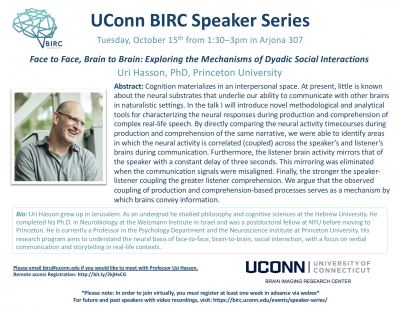
Talk: Uri Hasson, Princeton University
Uri Hasson, PhD
Princeton University
Tuesday, October 15th from 1:30-3:00 pm in Arjona 307
Abstract: Cognition materializes in an interpersonal space. At present, little is known about the neural substrates that underlie our ability to communicate with other brains in naturalistic settings. In the talk I will introduce novel methodological and analytical tools for characterizing the neural responses during production and comprehension of complex real-life speech. By directly comparing the neural activity timecourses during production and comprehension of the same narrative, we were able to identify areas in which the neural activity is correlated (coupled) across the speaker’s and listener’s brains during communication. Furthermore, the listener brain activity mirrors that of the speaker with a constant delay of three seconds. This mirroring was eliminated when the communication signals were misaligned. Finally, the stronger the speaker- listener coupling the greater listener comprehension. We argue that the observed coupling of production and comprehension-based processes serves as a mechanism by which brains convey information.
Bio: Uri Hasson grew up in Jerusalem. As an undergrad he studied philosophy and cognitive sciences at the Hebrew University. He completed his Ph.D. in Neurobiology at the Weizmann Institute in Israel and was a postdoctoral fellow at NYU before moving to Princeton. He is currently a Professor in the Psychology Department and the Neuroscience Institute at Princeton University. His research program aims to understand the neural basis of face-to-face, brain-to-brain, social interaction, with a focus on verbal communication and storytelling in real-life contexts.
**To view this talk remotely via Webex, please register here by October 8th**
Please email birc@uconn.edu if you are interested in meeting with a speaker. Click here to see the full BIRC Speaker Series schedule and access recordings of past talks.
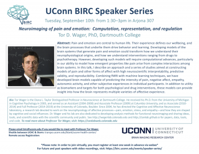
Talk: Tor Wager, Dartmouth College
Dartmouth College
Tuesday, September 10th from 1:30-3pm in Arjona 3o7
Abstract: Pain and emotion are central to human life. Their experience defines our wellbeing, and the brain processes that underlie them drive behavior and learning. Developing models of the brain systems that generate pain and emotion could transform how we understand their neurophysiological origins, and how we understand interventions ranging from drugs to psychotherapy. However, developing such models will require computational advances, particularly in our ability to model how emergent properties like pain arise from complex interactions among brain systems. In this talk, I describe an approach and a series of studies aimed at constructing models of pain and other forms of affect with high neuroscientific interpretability, predictive validity, and reproducibility. Combining fMRI with machine learning techniques, we have developed brain models capable of predicting the intensity of pain, negative affect, empathy, autonomic activity, and other subjective experiences in individual participants. In addition to utility as biomarkers and targets for both psychological and drug interventions, these models can provide insight into how the brain represents multiple varieties of affective experience.
Bio: Tor Wager is the Diana L. Taylor Distinguished Professor in Neuroscience at Dartmouth College. He received his Ph.D. from the University of Michigan in Cognitive Psychology in 2003, and served as an Assistant (2004-2008) and Associate Professor (2009) at Columbia University, and as Associate (2010-2014) and Full Professor (2014-2019) at the University of Colorado, Boulder. Since 2004, he has directed the Cognitive and Affective Neuroscience laboratory, a research lab devoted to work on the neurophysiology of affective processes—pain, emotion, stress, and empathy—and how they are shaped by cognitive and social influences. Dr. Wager and his lab are also dedicated to developing analysis methods for functional neuroimaging and sharing ideas, tools, and scientific data with the scientific community and public. See http://wagerlab.colorado.eduand http://canlab.github.iofor papers, data, tools, and code.
**To view this talk remotely via Webex, please register here by September 3rd**
Please email birc@uconn.edu if you are interested in meeting with a speaker. Click here to see the full BIRC Speaker Series schedule and access recordings of past talks.
Expanding Minds: BIRC Community Outreach
The UConn BIRC was recently featured in UConn Today for community outreach. The center hosted students from E.O. Smith High School for a tour of the facility and informational sessions led by faculty, staff, and graduate students.
For the original story in UConn Today and photographs, please click here.
Fumiko Hoeft Receives Eye-to-Eye Academic Excellence Award
Fumiko Hoeft, MD, PhD recently received an award from nonprofit organization Eye-to-Eye for her work with Stephanie Haft: Impact of mentoring on socio‐emotional and mental health outcomes of youth with learning disabilities and attention‐deficit hyperactivity disorder. The paper can be accessed here.
To learn more about Eye-to-Eye and their mission, visit their website.
InCHIP Virtual Meet ‘n’ Greet: UConn Brain Imaging Imaging Research Center
Dr. Fumiko Hoeft, Director of the UConn Brain Imaging Research Center (BIRC) shares information regarding the state-of-the-art equipment, methods, and training offered by BIRC and how the center supports both brain and whole-body imaging and research across the life span in addition to a range of clinical and nonclinical populations. She also covers BIRC equipment that can be used for research purposes includes MRI/fMRI scanner, TMS, tDCS/tACS, and EEG.
Also, watch to learn more about a $30,000 seed grant opportunity that InCHIP and BIRC are co-sponsoring!
Click here to watch the full InCHIP Virtual Meet ‘n’ Greet BIRC Seminar
Professors Myers and Eigsti Receive Five Year Training Grant
UConn BIRC Trailblazer Award Announced
Issue Date
December 19, 2018
Background
Since the opening of the University of Connecticut (UConn) Brain Imaging Research Center (BIRC) in June 2015, there has been an increase and diversification of user-base, neuroimaging-related extramural grants, and neuroimaging expertise of students and faculty. However, there is still room for greater utilization of BIRC, which presents opportunities for BIRC to offer the resources to perform high-profile and neuroimaging-intensive research that other fully occupied imaging centers cannot offer.
Objective
The BIRC Trailblazer Award was created to allow research teams to perform cutting-edge research and/or perform research that will benefit the BIRC community at-large. The objective of the 2019 BIRC Trailblazer Award is to fund: (1) high-risk high-reward projects with exceptional innovation that lead to raising the visibility of UConn, College of Liberal Arts and Sciences (CLAS) and BIRC; and/or (2) projects that will benefit the BIRC community at-large (e.g. methods development). The project is intended to lead to high-profile peer-review publications, release of a public database, and/or work that is cited and utilized by large-number of UConn researchers in their grants and manuscripts. The project should also lead to large-scale and high-profile extramural grant applications shortly after the end of the funding period.
Clinical MRI scans now being offered at BIRC
UConn Health Patients Can Now Get MRIs at UConn in Storrs
UConn Health patients in eastern Connecticut will now be able to get MRI scans done in Storrs just as if they were at UConn Health in Farmington, thanks to a collaboration between doctors and researchers at the two campuses.
UConn’s Brain Imaging Research Center (BIRC) houses a powerful 3 Tesla Magnetic Resonance Imaging (MRI) scanner that was installed in 2015 and originally dedicated purely to research. The BIRC’s machine can take detailed pictures of fine structures in the brain. It can also do functional MRI, which shows subtle changes in blood flow in the brain as a person thinks. Medical details – tiny flecks of blood that might signal a concussion, or small injuries to the spine or extremities – also show up beautifully on the scanner. But the state had not previously licensed the BIRC’s machine to perform medical work. Doctors at UConn Health and researchers at the BIRC thought that should change.
“Soon after I started as chair, it became clear we had a long history of our UConn Husky athletes having scans done on the outside. But then their docs would bring the scans to us for a second read because they trusted us,” says Dr. Leo Wolansky, head of radiology at UConn Health. “It’s our moral obligation to take care of our own people,” but it was a lot of unpaid work too, he observes. Other patients in eastern Connecticut, including students, staff, and faculty, who wanted to use UConn Health doctors but worked and lived near Storrs also found it inconvenient to drive to Farmington for a scan. That meant a lot of UConn money leaving the institution, Wolansky adds, an undesirable phenomenon known as “leakage.”
So he began working with former BIRC scientific directors Inge-Marie Eigsti and Jay Rueckl, and more recently BIRC director Fumiko Hoeft, along with regulatory and business development staff at UConn Health, to get permission from the state to use the center’s machine for medical imaging. Then, with the help of UConn Health MRI technologist Brian Hausner, MRI service manager Elisa Medeiros installed protocols to run clinical scans. The picture archiving communication system team at UConn Health oversaw the setup of the hardware needed to transmit medical data securely from the BIRC, which is located in the Phillips Communication Sciences Building in Storrs, to UConn Health in Farmington.
It took months of work, but on Nov. 7, the BIRC scanned Clinical MRI Patient #1. The term “Clinical MRI Patient #1” had a double meaning. It referred to the first clinical scan chronologically, but it was also a HIPAA-compliant way to refer to the top executive at UConn, President Herbst, who agreed to be the “test case.” She has given permission to publish this information, and wants everyone to know she’s fine.
“The President’s support of the project was critical,” says Wolansky.
UConn Health doctors can now schedule MRIs for their patients at the BIRC in Storrs for Monday and Wednesday afternoons as easily as if they were going to the imaging center in Farmington. Urgent scans can be squeezed in at other times on a case-by-case basis. The BIRC capacity will free up some space at UConn Health, bringing new patients into the system, and is not expected to impact research done at the center at all.
“The biggest benefit is the integration between campuses. It’s a huge success for us to do this,” says Hoeft, the director of BIRC, noting that revenue from the scans will enhance the financial stability of the center.
Wolansky, who is based in Farmington, agrees. UConn’s Farmington and Storrs campuses function largely independently from one another, so this collaboration is based on goodwill, he says.
“They are UConn and we are UConn,” he adds. “Even though [patients] may be 40 minutes away by car, when we read the scans [at the imaging center in Farmington], it’s no different than if the patients were down the hall!”
This story was originally published in UConn Today